ProLife Medical® · Imaging centre, Bârlad

Medical imaging: MRI, CT and X-ray

Diagnostic examinations to investigate, characterise and monitor neurological, musculoskeletal, chest, abdominal and other conditions, according to the clinical indication and available examination protocols.

Strada Dr. Codrescu nr. 3, Bârlad Monday–Friday 07:30–20:00 · Closed on weekends

The map is not loaded automatically. If you choose to display it, Google Maps may receive technical data, including your IP address, and may use cookies. Cookie policy (RO).

Open in Google Maps ↗

Imaging examinations

Complementary methods for different diagnostic questions

MRI and CT provide different information about anatomy and disease. The medical team selects the anatomical region, examination method and any contrast-enhanced protocol according to the clinical question.

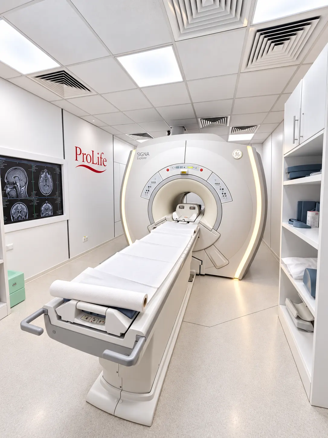
ProLife Medical MRI examination room with GE SIGNA Explorer
GE SIGNA Explorer · 1.5 Tesla MRI

MAGNETIC RESONANCE IMAGING

Multiplanar assessment of soft tissues and anatomical structures

MRI uses magnetic fields and radiofrequency pulses to produce high soft-tissue contrast without ionising radiation. T1- and T2-weighted imaging, fat suppression and diffusion sequences can support assessment when the protocol and clinical indication require them.

Clinical indications and applications
  • Neuroradiology: cerebrovascular disease, demyelinating lesions, intracranial masses, inflammatory changes and post-injury findings.
  • Spine: disc disease, herniated discs, spinal canal stenosis, nerve root compression, myelopathy and spinal cord abnormalities.
  • Musculoskeletal system: ligament, meniscus and tendon injuries, bone marrow oedema, cartilage and soft tissues.
  • Abdomen and pelvis: assessment of selected liver, pancreas, kidney or pelvic lesions under dedicated protocols.

These examples describe possible clinical uses of MRI. Please confirm the availability of specialist protocols at ProLife before booking.

MRI preparation and safety

Report pacemakers, defibrillators, neurostimulators, cochlear implants, aneurysm clips, implanted pumps and any metal fragments, especially in or near the eyes. MRI compatibility is specific to the exact device and its conditions of use. Bring the device documentation.

You must remain still, use hearing protection and notify reception in advance if you have claustrophobia. For MRI with gadolinium contrast, the centre requires recent urea and creatinine results, as explained above.
MRI service and detailed information (RO)
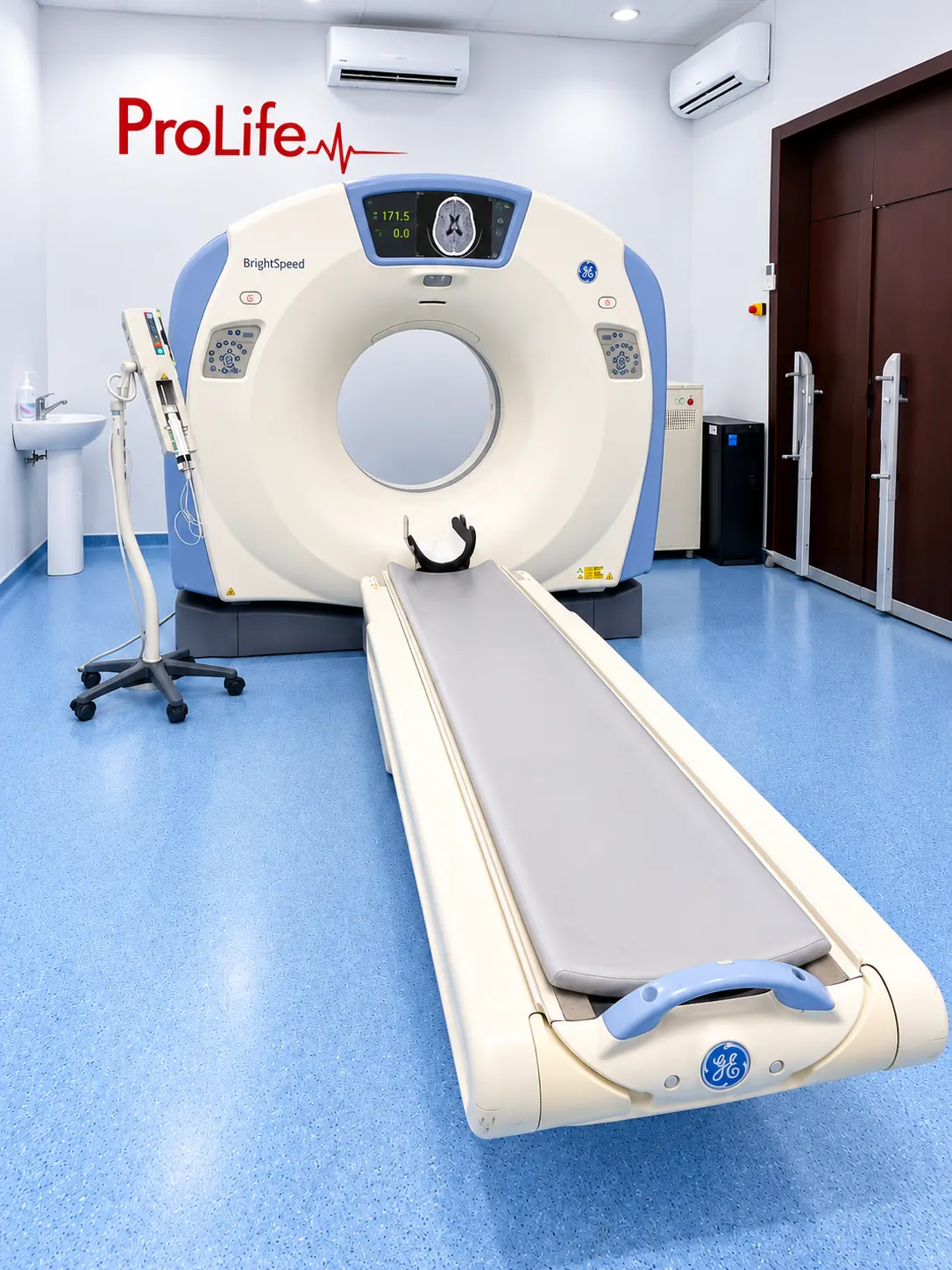
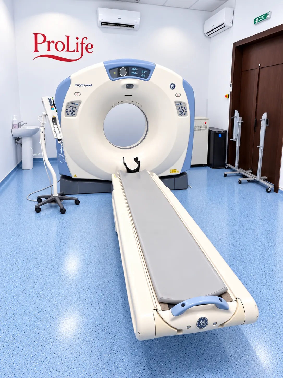
GE BrightSpeed CT scanner photographed at ProLife Medical
GE BrightSpeed · CT examination room photographed at ProLife

COMPUTED TOMOGRAPHY

Cross-sectional imaging of organs, bones and vascular structures

CT uses X-rays and computer reconstruction to image anatomical structures in axial sections and multiplanar views. It can characterise densities, anatomical relationships and the extent of certain abnormalities.

Clinical indications and applications
  • Head and facial structures: selected intracranial bleeding, post-traumatic changes, bones and sinuses, depending on protocol.
  • Chest: pulmonary nodules, interstitial lung changes, inflammation, pleura, mediastinum and selected oncology evaluations.
  • Abdomen and pelvis: inflammation, infections, liver, pancreatic and kidney abnormalities, urinary stones and oncology follow-up.
  • Bones and joints: complex fractures, bone abnormalities and detailed post-traumatic assessment.

CT angiography, multiphase studies and other specialised protocols require confirmation of technical availability and the medical indication before booking.

CT preparation and safety

CT involves medically justified and optimised exposure to ionising radiation. Tell the team if you are or might be pregnant. For iodinated contrast, the team considers previous reactions, kidney function, other conditions and medication. Do not stop treatment unless instructed by a clinician.

According to the current local protocol, urea and creatinine results must be no more than 48 hours old for contrast-enhanced CT; eGFR is requested when recommended by the physician. Non-contrast CT does not automatically require the same tests.

CT service and detailed information (RO)
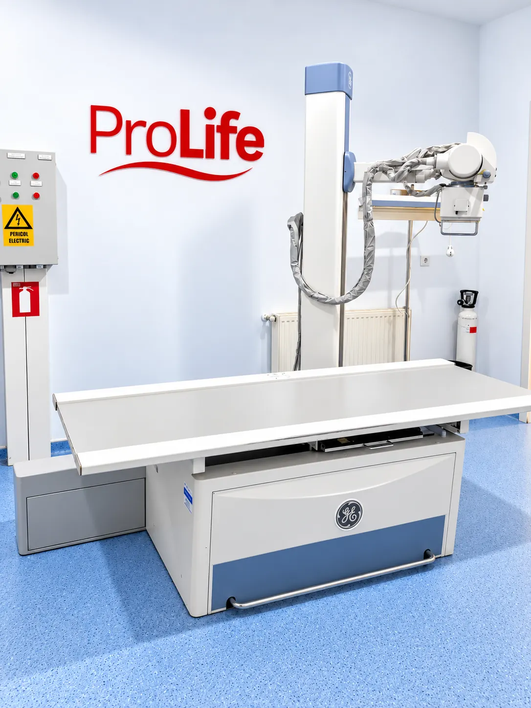
Conventional radiography equipment at ProLife Medical
Conventional X-ray equipment · GE
CONVENTIONAL RADIOGRAPHY

X-ray examinations

X-ray imaging is used to assess certain bones and structures in the chest. The required views, clinical indication and preparation depend on the body region. Please check availability with reception.

X-ray service (RO)

Patient journey

From clinical indication to the radiology report

01Indication and booking

Confirm the anatomical region, clinical question and whether contrast may be required.

02Screening and preparation

Review implants, previous contrast reactions, medical documents and requested laboratory tests.

03Image acquisition

The examination follows the selected protocol, with instructions from the radiographer.

04Radiology report

A radiologist describes the findings; your treating physician interprets them in clinical context.

Detailed clinical information

Guides by body region

The detailed guides listed below are currently available in Romanian. They explain common indications, preparations, documents and approximate examination times.

Practical patient information

Preparation, contrast agents and medical screening

Only follow the instructions relevant to your booked examination. The individual instructions from the imaging team take precedence.

01Preparing for your examinationTests, documents, food and timing

Contrast studies: local policy requires urea and creatinine results taken no more than 48 hours before MRI or CT with contrast; eGFR when recommended by a physician.

Documents: physician referral, ID, prior reports and images (preferably DICOM), relevant insurance documents, and information about implants or treatment.

Fasting, hydration and examination duration vary by protocol. Do not stop medication without clinical advice.

Full preparation guide (RO)
02Contrast agentsIodinated contrast for CT, gadolinium for MRI

Contrast use depends on the clinical question. Iodinated agents and gadolinium-based agents have different uses and safety profiles.

Report any previous contrast reaction, the agent involved if known, kidney disease, relevant medication, pregnancy or breastfeeding. The team makes an individual risk assessment.

Under the local protocol, urea and creatinine results must be no more than 48 hours old; eGFR is requested when medically indicated.

Contrast information (RO)
03Medical history and safety screeningImplants, contraindications, previous reactions

MRI: declare a pacemaker, defibrillator, cochlear implant, neurostimulator, vascular clip, implanted pump or possible metallic foreign bodies, especially intraocular ones. Device-specific conditions must be checked before entry into the MRI area.

Contrast: previous reactions, kidney function, medication and special circumstances require a separate assessment. Screening does not replace informed consent for the examination.

The detailed pre-examination questionnaire is currently available in Romanian and does not replace assessment by the radiologist.

Pre-examination questionnaire (RO)

ProLife Medical imaging centre

Equipment and examination rooms

Photographs of the imaging equipment and rooms. Please confirm the availability of specific investigations and protocols with reception before booking.

CT scanner at ProLife Medical imaging centre
GE BrightSpeed CT examination room
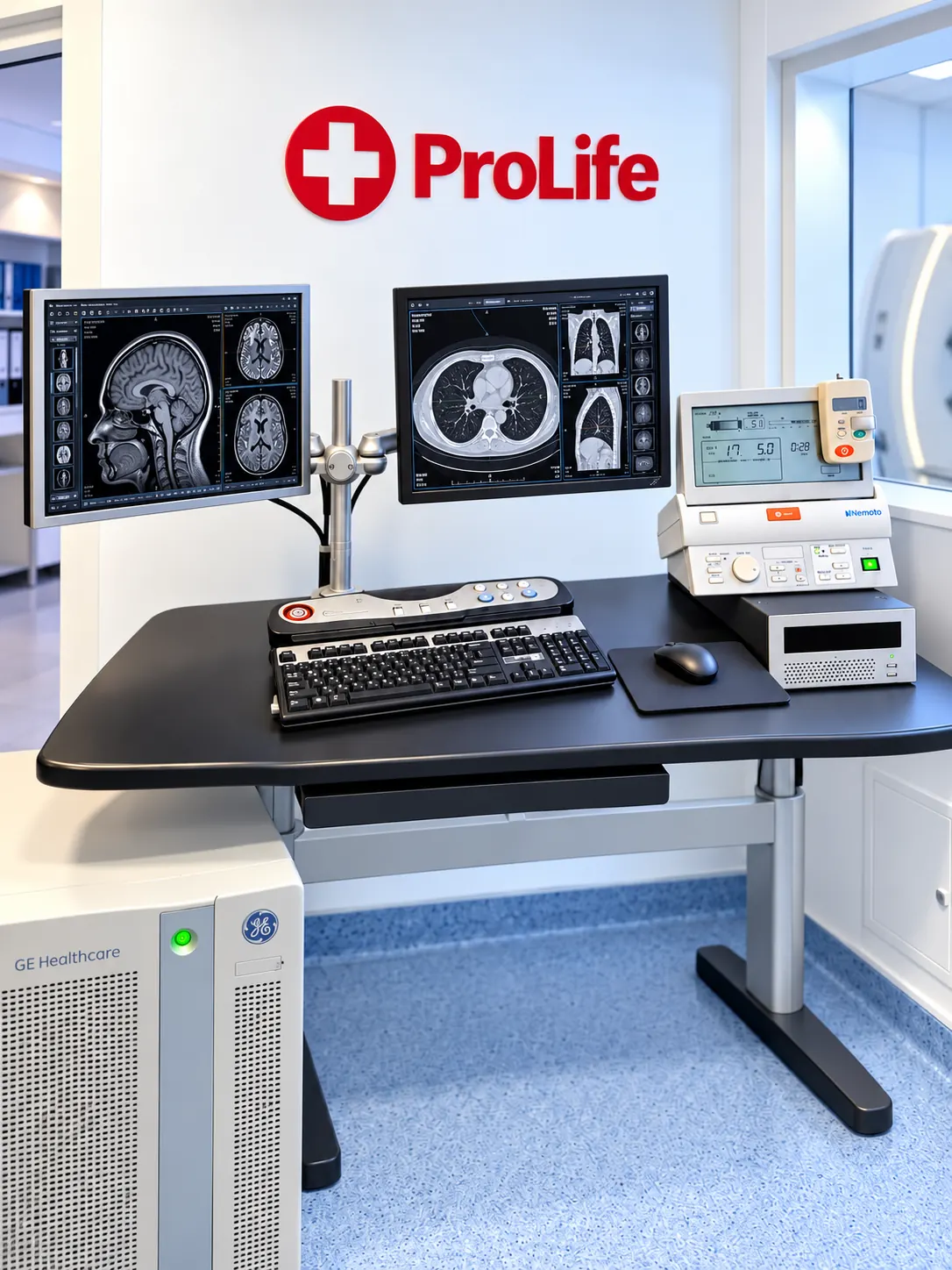
Imaging acquisition and reporting console at ProLife Medical
MRI and CT acquisition and display console
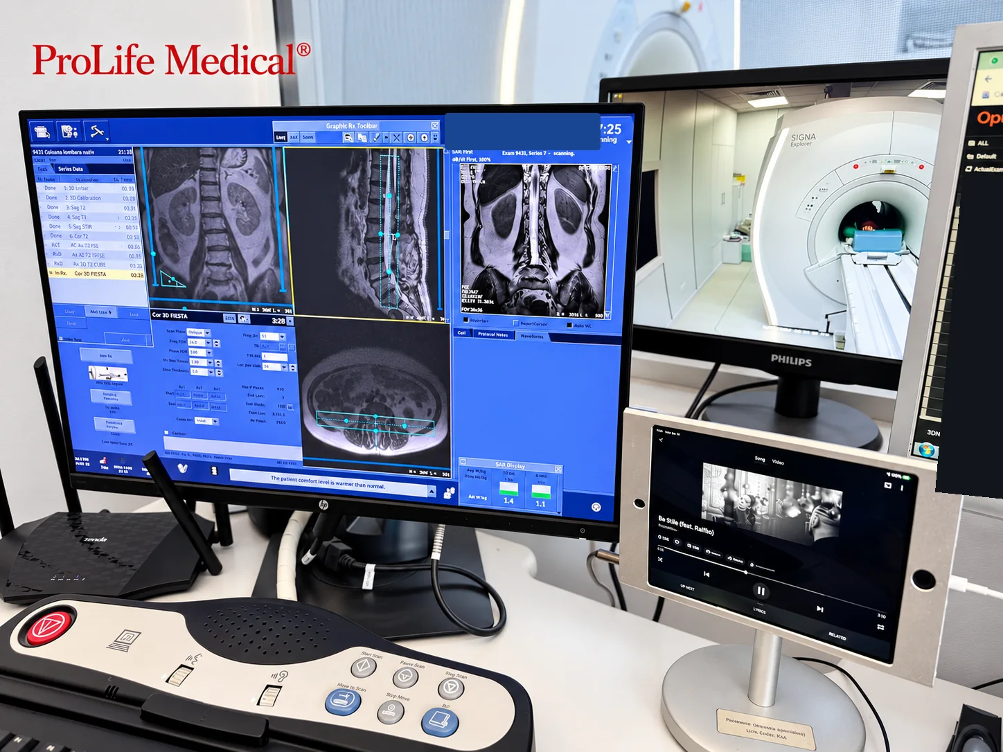
GE SIGNA Explorer MRI console at ProLife Medical
GE SIGNA Explorer MRI examination console

Appointments and documents

Private-pay and CAS Vaslui examinations

For examinations requested with a referral, reception checks the contracted service, required documents, eligibility and appointment availability for the requested period before confirming your visit.

Private-pay and reimbursed examinations may have different booking conditions and time slots. Please tell reception if you wish to use CAS coverage.

ProLife Medical imaging centre Strada Dr. Codrescu nr. 3, BârladMonday–Friday 07:30–20:00 · Closed on weekends Appointments and reception 0786 232 185

Patient questions

Preparing for MRI and CT: frequently asked questions

These answers describe the usual process. Final preparation depends on your examination and the assessment of the medical team.

Open MRI and CT questions and answers
01Medical documentsReferral, previous images and reports
02Urea and creatinineFor contrast, no more than 48 hours old under local protocol
03Safety screeningMRI implants, previous reactions, pregnancy and treatments
04Individual instructionsFood, timing and companions depend on the protocol

Before the examination

Booking, documents, laboratory tests and medication

Which documents and previous examinations should I bring?

Bring your ID, your physician's referral and any documents specified when booking. For reimbursed services, reception will clarify the correct referral type and eligibility documents.

Previous reports and imaging of the same region, ideally DICOM images, can be helpful. Bring the implant card or manufacturer documentation for any implanted devices before MRI.

Which laboratory tests are needed before MRI or CT with contrast?

ProLife's local protocol requires urea and creatinine results for contrast-enhanced MRI and CT, obtained no more than 48 hours before the examination. eGFR is requested if recommended by the physician.

These results do not replace the clinician's individual evaluation of renal function, previous reactions and whether contrast is appropriate. Non-contrast examinations do not automatically require the same tests.

Must I fast or stop medication?

Preparation differs between brain MRI, abdominal MRI, chest CT and abdominal or pelvic CT. Food, fluids and oral contrast instructions depend on the protocol. Do not stop metformin, anticoagulants or other medication on your own. Tell the team what you take.

How do I book an examination covered by CAS?

Tell reception that you require an examination under CAS Vaslui. Staff will verify the investigation, referral, contracted services, eligibility documents and available appointment dates. See CAS appointment information.

Safety and contraindications

Implants, contrast agents, pregnancy and radiation

Can I have an MRI with a prosthesis, stent or implanted device?

It depends on the exact device and its approved MRI conditions. Tell the team about pacemakers, defibrillators, cochlear implants, stimulators, pumps, surgical clips, prostheses and suspected metallic foreign bodies in the eyes. Bring model and manufacturer documents. Some devices are MR Conditional only under specific technical conditions.

What should I report about previous reactions or kidney disease?

Describe any previous reaction to iodinated CT contrast or gadolinium-based MRI contrast, the agent if known, symptoms and treatment. A general statement of 'iodine allergy' does not characterise a previous contrast reaction.

Inform the team about kidney disease, dialysis, transplantation, acute kidney injury and relevant medication. Recent laboratory results do not replace individual medical risk assessment.

What if I am pregnant or breastfeeding?

Report a confirmed or possible pregnancy before CT or MRI. With CT, ionising radiation exposure must be justified; with MRI, especially if gadolinium is considered, the physician weighs benefits, risks and alternatives.

For commonly used contrast agents, routine interruption of breastfeeding is generally not recommended by professional guidance, but tell the clinician which agent is planned and any special medical circumstances.

Does CT expose me to radiation, and how is the right test chosen?

CT uses X-rays; MRI does not use ionising radiation. The choice depends on the body region and the clinical question. CT is often useful for lungs, bones and certain acute conditions; MRI can provide more detail for many soft tissues and neurological structures. CT examinations must be justified, with exposure optimised for the diagnostic task.

I have claustrophobia or cannot remain still. Can I have MRI?

Tell reception before booking. MRI involves remaining still and produces loud noises; staff will explain communication and hearing protection. Additional arrangements may be needed for severe anxiety or pain. Do not assume sedation is available at the imaging centre; any such arrangements must be confirmed individually.

During and after the examination

Duration, assistance, monitoring and results

How long does MRI or CT take, and when should I arrive?

Duration depends on the number of regions, protocol and contrast. An MRI of one region commonly takes around 20–45 minutes, although some protocols take longer. CT acquisition is generally quicker, but preparation and checks can extend the overall visit. Reception confirms your arrival time and expected duration.

What will I feel, and how can I communicate with staff?

For MRI, you lie on the table and hear rhythmic scanning sounds; hearing protection is used. For CT, the table passes through the scanner ring, and you may be asked to hold your breath. Contrast injection can cause transient warmth or an unusual taste. Tell staff promptly about significant discomfort or unusual symptoms.

When do I need someone to accompany me?

Tell reception if the patient is a child, has reduced mobility, needs transfer assistance or may have difficulty cooperating. The centre will explain access and assistance requirements. Anyone entering the controlled MRI area requires magnetic safety screening. Sedation, if arranged and medically indicated, has its own accompanying and driving restrictions.

What should I watch for after receiving a contrast agent?

Follow individual observation and hydration instructions. Notify staff about injection-site pain or swelling, rash, itching or unusual symptoms. Breathing difficulty, facial or throat swelling, fainting or other severe symptoms require emergency medical assistance; outside the centre, call 112.

Who interprets my images and how do I obtain the report?

A radiology and medical imaging specialist interprets the images and prepares the radiology report. Reception explains when and how your report and, when applicable, images will be released. Your treating physician places the findings in clinical context and advises on treatment.

Have a specific medical concern?

Contact reception before visiting to clarify the required preparation, safety screening and documents.

Professional references

RadiologyInfo / ACR–RSNA: MRI preparation · RadiologyInfo / ACR–RSNA: CT · American College of Radiology: Contrast Manual

These explanations are educational. The medical indication, contrast administration and preparation are decided according to the clinical assessment and local protocol.

Your feedback helps us improve

The anonymous eight-question satisfaction survey is currently available in Romanian.

Patient satisfaction survey (RO)
Scroll to Top